Diagram Of Liver Pancreas And Gallbladder - Figure 34 10 Pancreas Liver And Gallbladder Diagram Quizlet - The pancreas delivers the digestive juice to the small intestine through small tubes called ducts.
Diagram Of Liver Pancreas And Gallbladder - Figure 34 10 Pancreas Liver And Gallbladder Diagram Quizlet - The pancreas delivers the digestive juice to the small intestine through small tubes called ducts.. The liver releases bile secretions which emulsify fats and enhances the activity of pancreatic and intestinal lipases. Human liver, gallbladder, pancreas anatomy vector. Secretes pancreatic juice it is the most important since ti can break down all three major kinds of food contains sodium bicarbonate which is an alkaline substance that. Sketch and label a diagram of the digestive system and describe it in your own words. Name the 3 components of the liver?
This results in distention/pain of hypochondrium and chest. Sketch and label a diagram of the digestive system and describe it in your own words. The liver releases bile secretions which emulsify fats and enhances the activity of pancreatic and intestinal lipases. Where is the pancreas located? Hepatocytes, bile caniculi and hepatic sinusoids.
Digsys J Liver Gall Bladder Pancreas Anatomy Youtube from i.ytimg.com It joins with the mesenteric vein inside the pancreas. It consists of bile salts, electrolytes (dissolved charged particles. Name the 3 components of the liver? State the main digestive roles of the liver, pancreas, and gallbladder identify three main features of liver histology that are critical to its function.on the activities of three accessory digestive organs: The liver, gallbladder, and pancreas are like big glands that sprouted from the digestive tract. It stores bile, a fluid made by your liver to. The remaining tissue consists of endocrine cells called. The liver is around the size of an american football at about 16 cm.
Pancreas, gallbladder and biliary tree.
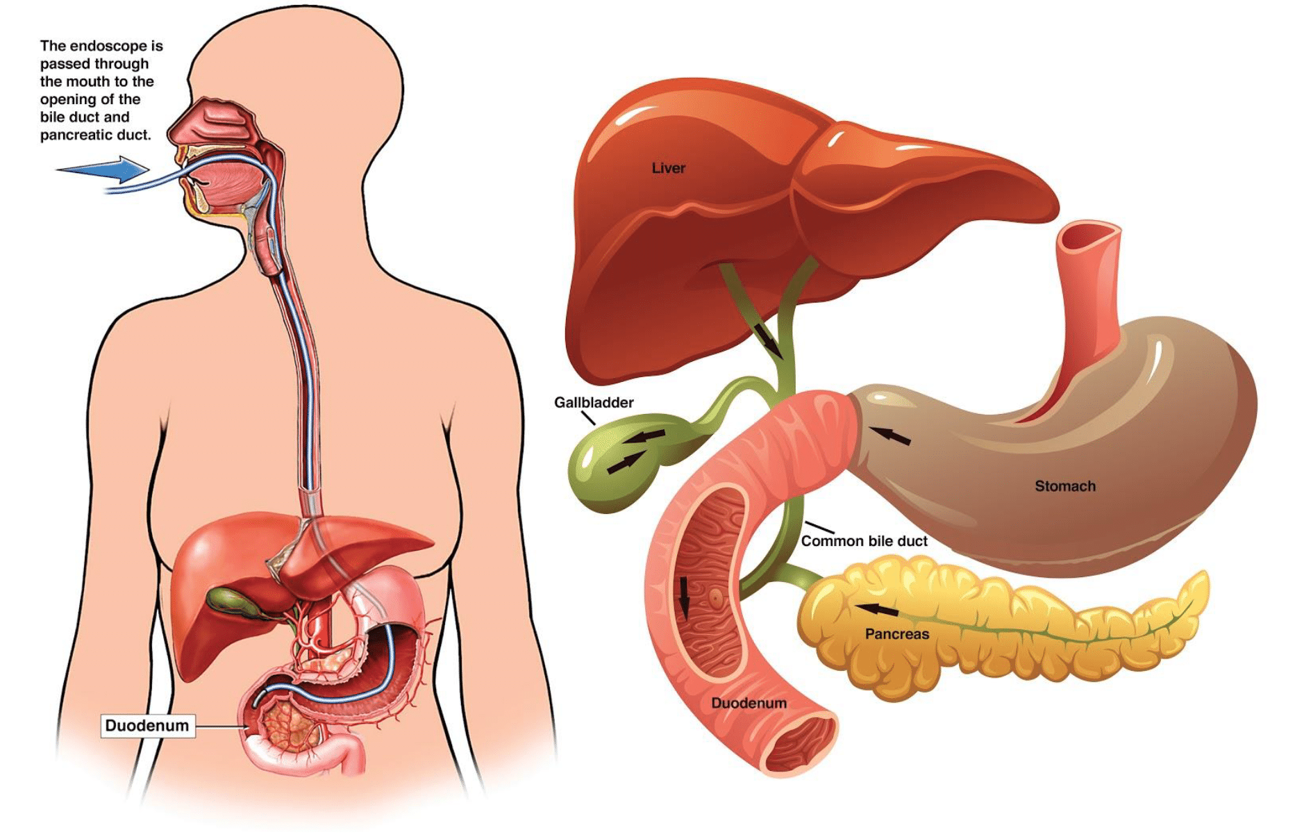
Controls all the other organs of the body and ensures they work together as a team. Advertisements help pay for this website. Name the 3 components of the liver? The liver, pancreas, and gallbladder. Endoscopic retrograde cholangiopancreatography (ercp) is an investigation used to view and if necessary biopsy the gallbladder, bile duct, pancreas, and pancreatic duct. To know the histological structure and major functions of the liver, gall bladder and pancreas. What is an exocrine gland? Location of liver in the human body. The liver, pancreas, and gallbladder are the solid organs of the digestive system. Your pancreas makes a digestive juice that has enzymes that break down carbohydrates, fats, and proteins. Then branches of the portal vein gives rise to venous sinusoids that pass between plates of liver cells. Oesophagus, gallbladder, liver, and pancreas. The hepatic portal system is designed to rid the.
Start studying pancreas, liver and gallbladder. The hepatic portal system is designed to rid the. Liver looks like the csf fluid if normal liver looks like spleen if abnormal. Included in the description of the. What is an exocrine gland?
Ercp Endoscopic Retrograde Cholangio Pancreatography Patient Information From Sages from www.sages.org The pancreas delivers the digestive juice to the small intestine through small tubes called ducts. The function of the gallbladder is to store the dilute bile it receives from the hepatic duct, concentrate it. It plays an essential role in converting the food we eat into fuel for the body's cells. Hepatocytes, bile caniculi and hepatic sinusoids. Start studying pancreas, liver and gallbladder. Identify the major types of enzymes and buffers present in pancreatic juice. Controls all the other organs of the body and ensures they work together as a team. Where is the pancreas located?
Secretions from the gall bladder and pancreas are released into the duodenum through a common structure, the hepatopancreatic ampulla, and the as part of the exocrine system, the pancreas secretes enzymes that work in tandem with bile from the liver and gallbladder to help break down.
Your pancreas makes a digestive juice that has enzymes that break down carbohydrates, fats, and proteins. Skip viragh center for pancreas cancer. To know the histology of the liver, including the hepatic lobule, hepatic acinus, hepatic parenchyma, portal area, and histological features of the vascular and biliary systems. The loss of pancreas tissue results in an inability to produce pancreatic fluids and thus to digest food properly, causing persistent flatulence and diarrhea. Liver looks like the csf fluid if normal liver looks like spleen if abnormal. Location of liver in the human body. The liver, gallbladder, and pancreas are like big glands that sprouted from the digestive tract. State the main digestive roles of the liver, pancreas, and gallbladder. What is an exocrine gland? The remaining tissue consists of endocrine cells called. The pancreas is an organ of the digestive system and endocrine system of vertebrates. Where is the pancreas located? It stores bile, a fluid made by your liver to.
Branch through ct and empty into the sinusoids (o2 rich. Gi cancers commonly metastasise to the liver, as venous blood returning from the bowel filters through the hepatic portal system first before rejoining the general circulation. To know the histology of the liver, including the hepatic lobule, hepatic acinus, hepatic parenchyma, portal area, and histological features of the vascular and biliary systems. Your pancreas makes a digestive juice that has enzymes that break down carbohydrates, fats, and proteins. Start studying pancreas, liver and gallbladder.
Untitled Document from eduspace.free.fr These organs work together to produce and a hollow muscular organ about the size of 2 closed fists, the stomach is located inferior to the diaphragm and lateral to the liver on the left side. Brain trachea (windpipe) lungs heart liver stomach spleen pancreas gallbladder kidneys bladder small intestines large intestines appendix. Start studying pancreas, liver and gallbladder. They are secretory glands that are associated with the alimentary system. Identify the major types of enzymes and buffers present in pancreatic juice. Controls all the other organs of the body and ensures they work together as a team. The liver releases bile secretions which emulsify fats and enhances the activity of pancreatic and intestinal lipases. The main pancreatic duct connects the pancreas with the small intestine.
Controls all the other organs of the body and ensures they work together as a team.
It stores bile, a fluid made by your liver to. State the main digestive roles of the liver, pancreas, and gallbladder identify three main features of liver histology that are critical to its function.on the activities of three accessory digestive organs: Advertisements help pay for this website. Almost all of the pancreas (95%) consists of exocrine tissue that produces pancreatic enzymes for digestion. The liver is around the size of an american football at about 16 cm. Where is the pancreas located? (see also overview of the liver and gallbladder.) bile is a greenish yellow, thick, sticky fluid. Sketch and label a diagram of the digestive system and describe it in your own words. Development and function of the pancreatic ducts: The liver, pancreas, and gallbladder. The loss of pancreas tissue results in an inability to produce pancreatic fluids and thus to digest food properly, causing persistent flatulence and diarrhea. 5 circulation to and from liver where does blood inferior side of liver at the porta hepatis where do the portal vein and hepatic artery empty? Oesophagus, gallbladder, liver, and pancreas.
Start studying pancreas, liver and gallbladder diagram of liver. The remaining tissue consists of endocrine cells called.